
")

Working group - a Portrait:
Experimental Cell- and Tissue Technology
The working group molecular haemostaseology* is a standalone institution of the University Hospital of Jena. The main topic of research is atherosclerosis and associated diseases such as strokes and heart attacks. Nearly 50 percent of all illness-related deaths in western society are attributed to atherosclerosis.
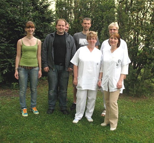
From left to right: Elisabeth Weiß (intern), Dr. Sandy Mosig (director of research group), outside lecturer Harald Funke (director of working group), Dr. Knut Rennert, Maria Franke (technical assistant), Silke Nossmann (technical assistant), Margot Voigt (technical assistant).
Photo: Rainer Spanbroeck
The research group Experimental Cell and Tissue Technology, featured here was set up as a part of this working group. The research group develops alternative investigative methods which aim to reduce animal use in biomedical research. They have been working for a number of years on the generation of artificial human blood vessel tissue. By applying modern tissue engineering, they seek to simulate the conditions within a living organism (in vivo) in laboratory surroundings (in vitro). Specifically, they use newly developed concepts of regenerative medicine for generating and simulating human organs (organoids). On the basis of their knowledge of processes of human pathogenetic processes and their therapy, they develop methods for realistic in vitro modelling and animal replacement.
The research group consists of three technical medical assistants, a doctoral student and graduate scientists. At present they are working on three particularly important projects:
1. Generation of artificial human blood vessels as a model for the investigation of the pathogeneses of atherosclerosis and sepsis
Ethical concerns regarding animal experiments are an important reason why the research group develops alternatives to animal models. Further, on account of the physiological differences between animals and humans, the results from animal experiments are not transferable to humans.
Often the organ preparation of killed animals only allows the use of their structures for a short time and therefore the course of disease can not be studied for more than a very brief period. Therefore the scientists, together with the industrial partners AVISO GmbH and Polymet e. V., and with the support of Zeiss Microimaging GmbH, are developing a three-dimensional flow chamber system. With this system and the help of appropriate optical methods, the adhesion and migratory behaviour of human cells in vitro and in real time can be observed.
In addition, important biological environmental conditions can be controlled and modified and the behavioural reaction of different cells of the blood vessel system can be studied. The project is supported by the Thuringian Ministry of Economics, Employment and Technology with funding from the EU and the State of Thuringia.
2. Development of a hepatic tissue model for the investigation of the infection process of human pathogen based on Francisella tularensis (rabbit fever)
In collaboration with Dr. Alejandro Soto-Gutierrez of the University of Pittsburgh/USA, and the Institute of Bacterial Infections and Zoonoses (IBIZ) of the National Research Institute for Animal Health (FLI) in Jena, the first artificial liver tissues, which are to serve as infection and pathogenesis models for zoonosis pathogens such as Francisella tularensis, were developed and tested.
Francisella tularensis is a gram-negative bacterium which causes tularaemia, a disease often lethal for rodents and transmissible to humans. Little is currently known about the infection mechanism and the course of tularaemia of humans. Likewise, the role of this monocytic persistent pathogen in the mediation of liver diseases is not yet fully understood. So far, studies of infection behaviour of Francisella have primarily been based on conventional monocellular cultures and on animal models. The development of an infection model of strains of Francisella should allow a better understanding of infection mechanisms und contribute to a sustainable avoidance of animal experiments in infection research.
3. Development of a microfluidic-supplied multi-organ system for reproducing metabolic interactions between liver, kidney and intestine
Within the research project, artificial human organ replicas of the liver will be integrated into microfluidic systems, which will be established as an in vitro model for pharmacological and toxicological tests of active agents. As well as liver tissue, other organ replicas such as kidneys and intestines are to be integrated. In this manner, nearly the whole human metabolic situation can be reproduced in vitro. Many toxins activated by liver tissue are not immediately toxic, but rather only develop their toxic effect s in neighbouring organs. By taking multi-organ interactions into consideration, peripheral toxic substances should also be ascertainable via in vitro tests. The development of multi-organ systems should contribute to a reduction in the amount of necessary animal experiments in pharmaceutical research and development.
Promising project already concluded: the "CellCelector"
An important and interesting project has just been concluded, the further development of the CellCelector of the company AVISO GmbH (now ALS GmbH). The CellCelector is a device initially used for single cell isolation of rare cells such as stem cells. The further development of the CellCelector enables the detection of different cell types from cell mixtures such as blood and tissue by marking them with fluorescence-conjugated antibodies. The cell types are registered via automated microscopy and harvested with a robotic system. The whole procedure takes place in a fully air-conditioned and sterile enclosure. This enables a non-damaging isolation of particularly sensitive cells such as monocytes and can be used for particularly sensitive analyses, e.g. single cell polymerase chain reaction).
The director of the research group, cell biologist Dr. rer. nat. Sandy Mosig, gave an interview to InVitroJobs on the situation and perspectives of the research work.
The young researcher Dr. rer. nat. Sandy Mosig graduated at the Biological and Phamaceutical Faculty of the University of Jena in 2007 and has since specialised in atherosclerosis. In his work he examines gene expression profiles of monocytes* and T cells* during the development of atherosclerosis in patients with a hereditary lipometabolic disorder (familial hypercholesterolaemia).
InVitroJobs:
Dr. Mosig, how must one imagine the inflammatory response which causes plaque destabilisation and can lead to atherosclerosis?
Dr. Sandy Mosig:
Due to increased deposits of blood lipids on the vessel wall, local inflammatory responses in the blood vessel tissue develop. This causes a migration of immune cells from the blood stream into the vessel. Monocytes, an early stage of macrophages circulating in the blood, pick up the modified blood lipids. In the case of larger fat deposits in the blood vessel, there lipid-rich cells develop, also called foam cells because the fat droplets in the cells look like foam under the microscope. During the further course of the illness, these cells die, and an intensified inflammatory reaction and the formation of an atherosclerotic plaque ensue, this plaques comprising dead cells and modified fat deposits.
Should the plaque break up and enter the blood stream, a thrombus forms, which lead to an acute thrombotic occlusion, accompanied by a shortage of oxygen in the heart or brain. This vascular occlusion can result in a heart attack or stroke.
InVitroJobs:
What is die significance of monocytes/macrophages? What kind of recent research findings exists about the role of different monocyte subpopulations during development of atherosclerosis?
Dr. Sandy Mosig:
atherosclerosis-associated diseases such as heart attack or stroke are responsible for 50 percent of all deaths in western society. Monocyte-mediated inflammatory processes play an essential role in the formation of atherosclerosis. Current hypotheses on the development of this disease assume that modified low density lipoproteins (mLDL) are involved in starting the inflammation cascade after accumulating in the extracellular matrix (ECM)* of the blood vessel between intima* and media*. Within the ECM, it comes to further modification processes caused by numerous different enzymes, which leads to the activation of endothelial cells within the blood vessel. Expression of surface proteins on the activated endothelial cells facilitates first a recruiting of monocytes are recruited from the blood stream, then a migration of these cells into the blood vessel tissue. Within the vascular wall, the monocytes differentiate into macrophages, incorporating considerable amounts of enriched mLDL. Thus the accumulation of lipid rich macrophages (foam cells) typical to atherosclerosis takes place within the vessel. This means that monocytes are involved in the onset of the disease. Our working group is looking into the function of the individual subtypes with regard to the development of atherosclerosis. Our work indicates that both sub-populations could have specific functions in this context.
InVitroJobs:
How many types of three-dimensional tissue models are there, and what are their intended areas of application?
Dr. Sandy Mosig:
For the pathogenesis of numerous diseases, the interactions between the different parts of the blood system and the layer of endothelial cells of the blood vessel are determining factors. Complex interactions of the different parts of the blood vessels regulate adhesion, transmigration and differentiation of the migrated leucocytes. Currently available in vitro investigative models can not completely reproduce the in vivo situation. For this reason, animal models, especially mouse or rat models, are still the “gold standard” for studies of pathophysiological processes in vascular systems. Due to differences between the human physiology and the physiology of the animal model, some of these fundamental difference, the transferability of results is often inadequate.
It is therefore our goal to produce artificial human blood vessel tissue and to integrate it into perfusable micro-chamber systems. In order to reproduce the functions of human blood vessel tissue as realistically as possible, it is necessary to take the three-dimensional structure of the corresponding in vivo situation into account when creating artificial tissue. By producing such heterogeneous complex structures, it is possible to create reliable pathogenesis models and use these in pharmacological and toxicological research beyond the scope of existing methods. We seek to gain an understanding of adhesion and transmigration behaviour of immune cells in a state of perfusion on or in the blood vessel tissue by using fluorescence-based cell-marking methods. The use of microfluidic technique creates a basis for a standardisable and cost-efficient application of tissue models in pathogenesis research. The small magnitude of the artificial blood vessel tissue and the comparably low volume required mean that this method can be used for testing substances are cost-intensive in production.
A functional in vitro pathogenesis model of the arterial wall on a microfluidic basis is an attractive alternative to animal experiments for vascular medicine, both from an ethical and a scientific point of view.
InVitroJobs:
Which “complex environmental control elements” are additionally simulated as described on your homepage? What is the current stage of development of your models (pre-validation/validation)?
Dr. Sandy Mosig:
We are developing control elements for monitoring partial oxygen and carbon dioxide levels in the ambient atmosphere. Humidity and temperature (in nutrient media and ambient atmosphere) are also controlled. We plan to produce standardised and reproducible tissue that we can integrate into the flow chamber system.
InVitroJobs:
What other areas of application could the flow chamber system have?
Dr. Sandy Mosig:
The flow chamber system is also intended for use as a pathogenesis model in sepsis research. In the clinical picture of sepsis, the interaction between immune cells and the blood vessel is a central aspect of pathogenesis. Generally, sepsis has been understood as a clinical symptom which can be reduced to an overacting systemic immune reaction based on a bacterial infection. Each year, about 200 new cases per 100 000 inhabitants are recorded (1). This makes sepsis about as common as cardiovascular diseases. With a hospital mortality of 54 percent, it is the main cause of mortality in intensive care units and incurs treatment costs in excess of 1.7 billion euros in Germany alone. The clinical symptoms of sepsis are not caused directly by invasive microorganisms, but rather an onset of hypertension*, coagulopathy* and multiple organ dysfunction*, caused by a dysregulation of inflammatory processes in the patient. It is considered a verified fact that systemic inflammatory reactions due to sepsis lead to the activation of the coagulation system and simultaneously to an inhibition of anticoagulant processes* and to fibrinolysis*. Even though coagulation and fibrin deposition represent essential mechanisms of host response to a microbial infection, an overshooting response leads to a situation in which the coagulation itself contributes to the course of the disease by means of microvascular thrombosis and multiple organ dysfunction.
One of the most important characteristics of sepsis is the microvascular dysfunction* of activated endothelial cells. Components of bacterial cells walls, e. g. lipopolysaccharides (LPS), lead to an activation of pattern recognition receptors (PRR)* on the surface of endothelial cells and mediate the adhesion and migration of leucocytes. Leukocyte migration leads to a release of a wide variety of cytokines*, chemokines* and components of the coagulation system. In the further course, a complex and yet not fully understood interaction between the immune system components (granulocytes, monocytes and T cells) and the blood vessel (endothelial cells, smooth muscle cells and connective tissue cells) takes place. One of the results of these interactions is a cytoplasmatic bulge, a contraction of endothelial cells and a displacement of the endothelial cell layer, followed by an amplification of the inflammatory response due to increasing migration of thrombocytes and leucocytes into the damaged vessel.
The use of artificial blood vessel tissues in pharmacological and toxicological research beyond an application as a pathogenesis model is conceivable and envisaged. Hereby applications in the testing and approval of substance have priority.
InVitroJobs:
Is your group working on any other developments for replacing animal testing?
Dr. Sandy Mosig:
Further projects are planned.
InVitroJobs:
You work in collaboration with Analytik Jena AG, ALS GmbH, Jenpolymers Ltd,, Polymet e. V. and Carls Zeiss Microimaging GmbH, to name a few. Was it easy to get them enthusiastic about your innovations?
Dr. Sandy Mosig:
All our collaborative partners showed a high degree of interest in developing animal-free testing methods for medical research. Our experience of collaborative work has been positive in every respect.
InVitroJobs:
Do you receive enough support for your project work? What is the situation on the “research funds marketplace” like?
Dr. Sandy Mosig:
The situation on the “research funds marketplace” is highly competitive, as in many other areas of research. We received special support from the state of Thuringia and the Thuringian Ministry of Economy, Labour and Technology.
InVitroJobs:
What motivates young students/graduates? Are they interested in replacement methods? Is there great demand? What are the training prerequisites for graduates/young scientists? Must more be done for education in the area of replacement methods?
Dr. Sandy Mosig:
In our experience, students are highly interest in alternative methods for replacing animal experiments. Nonetheless, in our opinion the fundamental ideas of such developments should be more intensively introduced in education. Together with Prof. Dr. Stefan Lorkowski from the Institute of Nutritional Research of the Friedrich Schiller University Jena, we intend to prepare such offers for students.
InVitroJobs:
Dr. Mosig, thank you for the interview.
Overview of the relevant cooperation partners in the area of animal-free research
a) Industry
Analytik Jena AG
microfluidic ChipShop GmbH
Jenpolymer Ltd. & Co KG
arthrospec GmbH
Automated Lab Solutions ALS GmbH
GoBio Project “Intelligente siRNA für die therapeutische Anwendung” (spin-off Company from FSU Jena as Biano Science GmbH in 2011)
Research Center for Medicine technique and Biotechnology, Dep. Bioinstruments and Device Development GmbH
b) Academic Partners
Friedrich Loeffler Federal Institute for Bacterial Infections and Zoonosis, Prof. Neubauer
Technical University Ilmenau, Biomechatronic, Prof. Witte
Institute for Nutrition Sciences, working group Biochemistry of Nutrition, Prof. Stefan Lorkowski
Children’s Hospital of Pittsburgh, Center for Innovative Regenerative Therapies, University of Pittsburgh/USA, Dr. Alejandro Soto-Gutierrez
c) University Hospital of Jena
Institute of Pharmacology and Toxicology, PD Dr. Lupp
University Women's Hospital Jena, Working group Molecular Gynaecology, Prof. Dürst
Glossary:
Molecular Haemostaseology
Science of blood coagulation and its possible disorders investigated by molecular methods.
Monocytes
Precursor cells of macrophages (eating cells) which migrate into tissue, where they combat bacteria and activate lymphocytes (2).
Monocytes and macrophages naturally process LDL, an important part of lipid metabolism. Should the protein part of LDL be modified by acetylation, the oxidised LDL can not be processed and accumulates within the cells. Monocytes and macrophages thus degenerated are called foam cells.
Together with injured blood vessels, they are regarded as the main cause of atherosclerosis (3).
T cells
T cells are the centre of the T cell mediated immune system: They play an important role in defence against pathogens and able to control immunological processes, mediate to other cell groups and directly eliminate pathogens (4).
Extracellular Matrix (ECM)
Tissue comprises cells and an extracellular matrix. The ECM includes bones, teeth and tendons, but also the basal lamina, which separates the connective tissue from the epithelial layer (5). The ECM of a blood vessel contributes to blood vessel functions in many ways. It is the framework, and the complex network of elastic fibres can fulfil mechanical tasks well. The ECM supplies information to the vascular cells necessary to the regulation of the cell proliferation and differentiation. Molecules with in modulation of the blood vessel system can also be stored in the ECM (6).
Intima and Media
Inner and outer layer of the vascular wall of an artery (7)
Amplification
Multiplication of the genome or genetic information, in this case naturally produced in the cells (8).
Proteolysis
Degradation of proteins, in this case degradation of the extracellular matrix (9).
Hypertension
High blood pressure (10)
Coagulopathy
Coagulation disorder (11)
Multi Organ Dysfunction
Dysfunction of several organs (12)
Anticoagulant processes
Process for inhibition of blood clotting (13)
Fibrinolysis
Dissolution of a thrombus (14).
Microvascular dysfunction
Malfunction of vessels (15).
Pattern Recognition Receptors (PRR)
Proteins which can recognise pathogens according to special patterns (16)
Cytokines, chemokines
Cytokines are polypeptides which are secreted by endothelial and epithelial cells (17).
Chemokines are a sub-family of cytokines that respond to inflammation signals with strong chemotactic activity (18).
Sources:
(1) “Centre for Sepsis Control and Care” (CSCC), University Hospital of Jena (http://www.cscc.ukj.de/Sepsis.html)
(2) http://www.med4you.at/laborbefunde/lbef_mono.htm
(3) http://de.wikipedia.org/wiki/Monozyt#Monozyten-assoziierte_Krankheiten
(4) Frömmel, C. (o. J.): Expressionsanalyse des Killer-Inhibitory-Rezeptors CD158b auf CD8 T-Lymphozyten, die spezifisch für Humanes Cytomegalievirus sind. Dissertation, Universitätsmedizin der Charité Berlin. http://www.diss.fu-berlin.de/diss/servlets/MCRFileNodeServlet/FUDISS_derivate_000000002285/0_Gesamtdissertation.pdf;jsessionid=77FCB65AF4FB149462000C51261CEA99?hosts=
(5) http://www.uni-leipzig.de/~biophys/gallery/wahlfach_schiller/ecm_physiker_web.pdf
(6) Eble, J. A. & Niland, S. (2009): Die extrazelluläre Matrix der Blutgefäße. Current Pharmaceutial Design 15/12: 1385-1400 (16).
(7) http://www.gesundheits-lexikon.com/Vorsorge-Diagnostik/Herz-Kreislauf-Gefaesse/Intima-Media-Dicke-Messung.html
(8) http://de.wikipedia.org/wiki/Amplifikation_%28Genetik%29
(9) http://flexikon.doccheck.com/Proteolyse
(10) http://www.netdoktor.de/Krankheiten/Bluthochdruck/
(11) http://www.medizinfo.de/haematologie/blutungsneigung/koagulopathie.shtml
(12) http://sciencev1.orf.at/science/news/117557
(13) http://de.wikipedia.org/wiki/Antikoagulation
(14) http://www.herzberatung.de/blutgerinnungshemmung_fibrinolyse.html
(15) http://de.wikipedia.org/wiki/Multi-Dysfunktionalit%C3%A4t
(16) http://www.freidok.uni-freiburg.de/volltexte/5712/pdf/korrigierteVersion2008b.pdf
(17) Engelhardt, B. (o. J.): IMMUNOLOGIE II, Kapitel 12 – Zytokine. University of Bern. http://www.immunology.unibe.ch/wiki/Files/Kapitel_12_Immu_II_BE.pdf
(18) http://www.ipa.ruhr-uni-bochum.de/publik/info0103/chemokine.php
 Dr. rer. nat.